

Presacral Space
Author: Sarah Andiman, MD
Expert Reviewer: Marlene Corton, MD
Preferred English Term:
Presacral Space
Non-preferred Synonyms:
Retrorectal Space
Author: Sarah Andiman, MD
Expert Reviewer: Marlene Corton, MD
Preferred English Term:
Presacral Space
Non-preferred Synonyms:
Retrorectal Space
Click on an image to view a larger version along with its description.
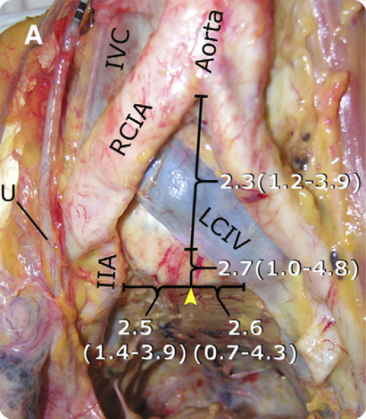
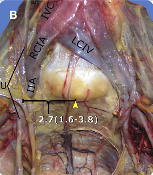

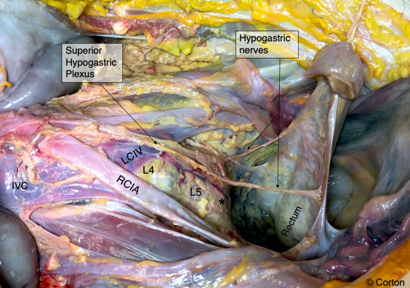
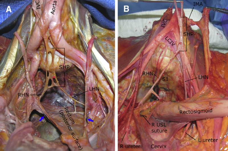
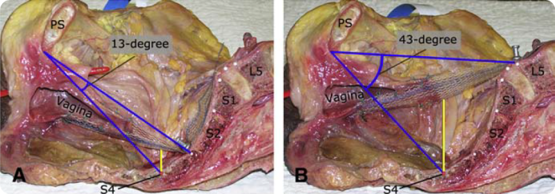
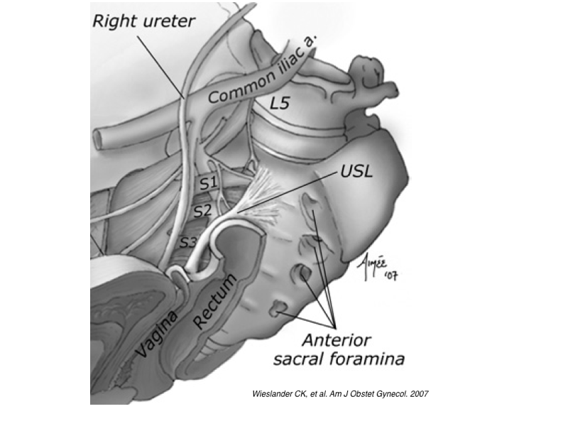
Anterior Longitudinal Ligament • Aortic Bifurcation • Common Iliac Artery • Common Iliac Vein • Hypogastric Nerve • Internal Iliac Artery • Internal Iliac Vein • Median Sacral Artery • Median Sacral Vein • Rectum • Sacral Promontory • Sacral Venous Plexus • Sacrum • Sigmoid Colon • Superior Hypogastric Plexus • Ureter
Balgobin S, Good MM, Dillon SJ, Corton MM. Lowest colpopexy sacral fixation point alters vaginal axis and cul-de-sac depth. Am J Obstet Gynecol. 2013 Jun;208(6):488. doi: 10.1016/j.ajog.2013.03.006. PMID: 23500452.
Florian-Rodriguez ME, Hamner JJ, Corton MM. First sacral nerve and anterior longitudinal ligament anatomy: clinical applications during sacrocolpopexy. Am J Obstet Gynecol. 2017 Nov;217(5):607.e1-607.e4. doi: 10.1016/j.ajog.2017.07.008. PMID: 28709584.
Good MM, Abele TA, Balgobin S, Schaffer JI, Slocum P, McIntire D, Corton MM. Preventing L5-S1 discitis associated with sacrocolpopexy. Obstet Gynecol. 2013 Feb;121(2 Pt 1):285-290. doi: 10.1097/AOG.0b013e31827c61de. PMID: 23344278.
Good MM, Abele TA, Balgobin S, Montoya TI, McIntire D, Corton MM. Vascular and ureteral anatomy relative to the midsacral promontory. Am J Obstet Gynecol. 2013 Jun;208(6):486.e1-7. doi: 10.1016/j.ajog.2013.02.039. PMID: 23454254.
Grimes CL, Tan-Kim J, Garfin SR, Nager CW. Sacral colpopexy followed by refractory Candida albicans osteomyelitis and discitis requiring extensive spinal surgery. Obstet Gynecol. 2012 Aug;120(2 Pt 2):464-468. doi: 10.1097/AOG.0b013e318256989e. PMID: 22825267.
Hare AM, Sawyer P, Hull S, Corton MM. Presacral Space Surgical Anatomy: Piriformis Fascia as Lateral Border. Urogynecology (Phila). 2025 Jun 1;31(6):583-589. doi: 10.1097/SPV.0000000000001681. PMID: 40168452.
Nygaard IE, McCreery R, Brubaker L, Connolly A, Cundiff G, Weber AM, Zyczynski H; Pelvic Floor Disorders Network. Abdominal sacrocolpopexy: a comprehensive review. Obstet Gynecol. 2004 Oct;104(4):805-23. doi: 10.1097/01.AOG.0000139514.90897.07. PMID: 15458906.
Ripperda CM, Jackson LA, Phelan JN, Carrick KS, Corton MM. Anatomic relationships of the pelvic autonomic nervous system in female cadavers: clinical applications to pelvic surgery. Am J Obstet Gynecol. 2017 Apr;216(4):388.e1-388.e7. doi: 10.1016/j.ajog.2016.12.002. PMID: 27956200.
Sato H, Kurita M, Abe H, Otsuka S, Tsukada S. Sagittal Double-Hump Deformity of the Lumbosacral Spine: An Anatomical Risk Factor for Surgical Landmark Obscuration During Laparoscopic Sacrocolpopexy. Neurourol Urodyn. 2026 Feb 9. doi: 10.1002/nau.70228. PMID: 41657298.
White AB, Carrick KS, Corton MM, McIntire DD, Word RA, Rahn DD, Wai CY. Optimal location and orientation of suture placement in abdominal sacrocolpopexy. Obstet Gynecol. 2009 May;113(5):1098-1103. doi: 10.1097/AOG.0b013e31819ec4ee. PMID: 19384126.
Wieslander CK, Rahn DD, McIntire DD, Marinis SI, Wai CY, Schaffer JI, Corton MM. Vascular anatomy of the presacral space in unembalmed female cadavers. Am J Obstet Gynecol. 2006 Dec;195(6):1736-41. doi: 10.1016/j.ajog.2006.07.045. PMID: 17132475.
Wieslander CK, Roshanravan SM, Wai CY, Schaffer JI, Corton MM. Uterosacral ligament suspension sutures: Anatomic relationships in unembalmed female cadavers. Am J Obstet Gynecol. 2007 Dec;197(6):672.e1-6. doi: 10.1016/j.ajog.2007.08.065. PMID: 18060977.
Knowing the relevant anatomy related to the abdominal sacrocolpopexy
SGS Video Library, Videofest 2014, Overview of presacral anatomy
Anatomic considerations of sacrocolpoperineopexy
SGS Video Library, Video Presentation 2013, Anatomy of presacral space
Robotic sacrocolpopexy
SGS Video Library, Video Presentation 2007, Robotic technique for sacrocolpopexy
You must have an account to view the terms.
Would you like unlimited access to all terms?
Request PAG Digital Library Access
Already a member? Click here to login.
