Levator Ani

Author: Terry White, MD
Editor: Caroline Cox, MD
Expert Reviewer: John O. L. DeLancey, MD

Preferred English Term: Levator ani
Preferred Latin Term: Musculi levator ani
Non-preferred Synonyms: Levator muscles, levator, levator ani complex, levator ani muscle, levator arch, levator plate, levator sling, pelvic floor muscles

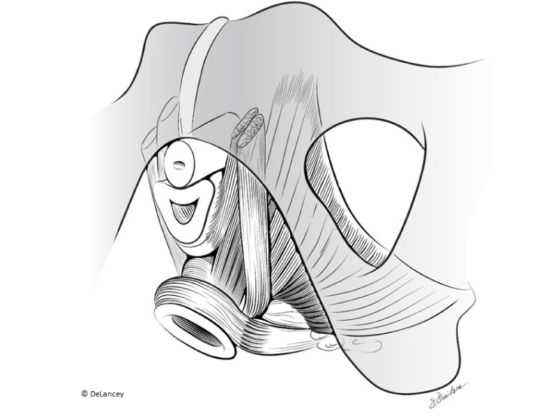
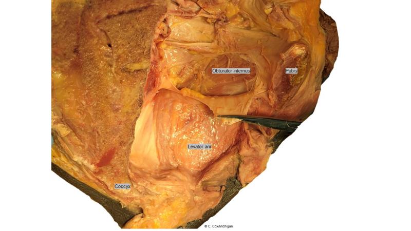
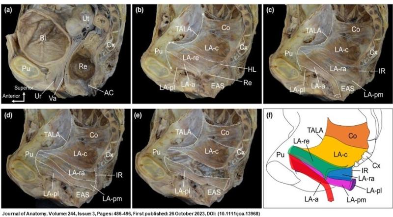
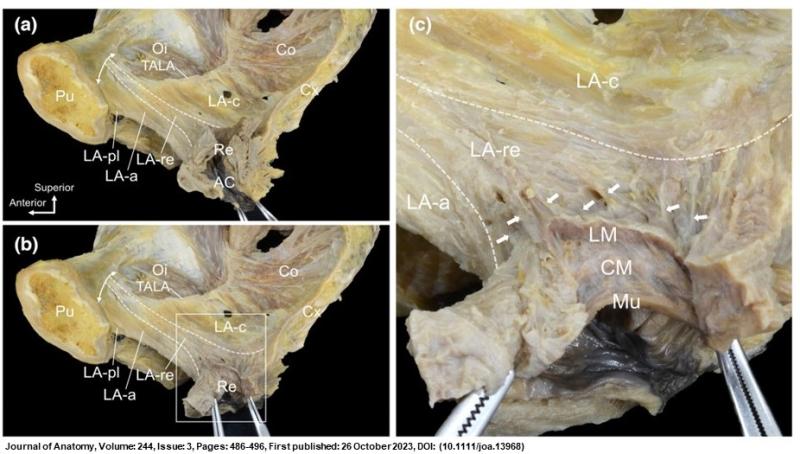
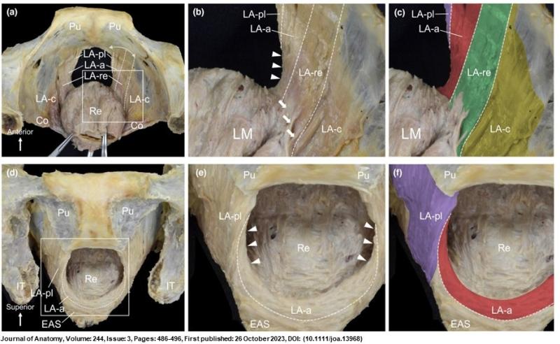
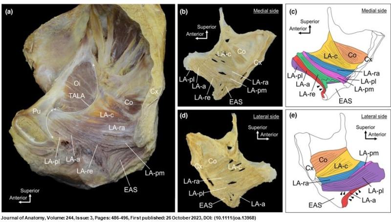
  • Composed of three collections of muscle fibers: pubococcygeus, puborectalis, and iliococcygeus.

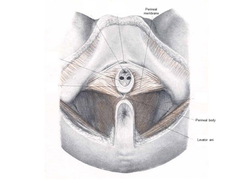
  • Musculature comprising significant component of the pelvic floor, supporting the pelvic organs.

    • Iliococcygeous acts as shelf while pubococcygeus and puborectalis loop around the hiatus with activation closing and opening the pelvic floor.

  • Resting tone helps to maintain closure of the rectum and vagina.

    • Acting reciprocally with the striated muscle of the anterior abdominal wall, functions to maintain the equilibrium of intra-abdominal pressure.

  • Relaxation facilitates elimination from the urinary and gastrointestinal tracts.

Clinical and Surgical Relevance

  • Muscular injury to the levator ani during pregnancy and vaginal birth can contribute to development of pelvic organ prolapse and stress urinary incontinence.

  • Levator ani is the focus of pelvic floor muscle training for various pelvic floor disorders.

  • Levator myalgia can be a cause of chronic pelvic pain.

Gross Anatomy

Click on an image to view a larger version along with its description.

Illustrations

Click on an image to view a larger version along with its description.

Videos

Introduction to levator ani anatomy in a cadaver dissection
Cadaver dissection exhibiting dynamic anatomy of the levator ani
Cadaver dissection highlighting direction of levator ani muscle fibers
Effect of levator ani contraction demonstarted in a standing cadaver
Levator Ani anatomy in 3D
MRI image scrolling from caudal to cranial direction in axial plane, Levator ani outlined in lavender
MRI image scrolling from posterior to anterior direction in coronal plane, Levator ani outlined in lavender

Recommended Resources

DeLancey JOL, Masteling M, Pipitone F, LaCross J, Mastrovito S, Ashton-Miller JA. Pelvic floor injury during vaginal birth is life-altering and preventable: what can we do about it? Am J Obstet Gynecol. 2024 Mar;230(3):279-294.e2. doi: 10.1016/j.ajog.2023.11.1253. Epub 2024 Jan 2. PMID: 38168908.

DeLancey JO, Mastrovito S, Masteling M, Horner W, Ashton-Miller JA, Chen L. A unified pelvic floor conceptual model for studying morphological changes with prolapse, age, and parity. Am J Obstet Gynecol. 2023 Nov 29:S0002-9378(23)02061-6. doi: 10.1016/j.ajog.2023.11.1247. Epub ahead of print. PMID: 38036167.

DeLancey JO, Pipitone F, Masteling M, Xie B, Ashton-Miller JA, Chen L. Functional Anatomy of Urogenital Hiatus Closure: the Perineal Complex Triad Hypothesis. Int Urogynecol J. 2024 Feb;35(2):441-449. doi: 10.1007/s00192-023-05708-w. Epub 2024 Jan 11. PMID: 38206338.

Muro S, Moue S, Akita K. Twisted orientation of the muscle bundles in the levator ani functional parts in women: Implications for pelvic floor support mechanism. J Anat. 2024 Mar;244(3):486-496. doi: 10.1111/joa.13968. Epub 2023 Oct 26. PMID: 37885272